
Brain CT Clinical Guide
ImagingGuide
27 articles
Abnormalities on computed tomography (CT) of the brain can be quite subtle, but half the battle is knowing what you’re looking for and where to find it. This guide will help you recognize common abnormalities on brain CT and how to identify when normal structures are absent. From traumatic brain injuries and strokes to hemorrhages, tumors, and seizures, you’ll gain the skills you need to avoid common pitfalls and maximize your ability to recognize subtle disease on brain CT.
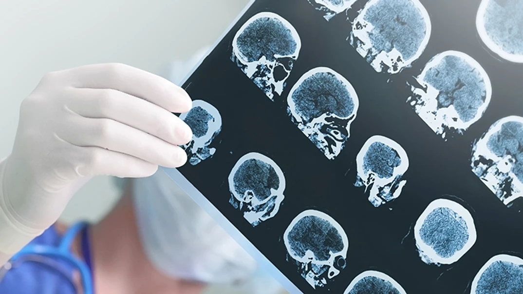
Abnormalities on computed tomography (CT) of the brain can be quite subtle, but half the battle is knowing what you’re looking for and where to find it. This guide will help you recognize common abnormalities on brain CT and how to identify when normal structures are absent. From traumatic brain injuries and strokes to hemorrhages, tumors, and seizures, you’ll gain the skills you need to avoid common pitfalls and maximize your ability to recognize subtle disease on brain CT.

Editors:Shelley Jacobs, PhD
Peer reviewers:Franz Wiesbauer, MD MPH Internist
Last update 16th Aug 2024
Chapter 1
The clinical essentials of brain computed tomography (CT)
To competently read a computed tomography (CT) brain scan, you need to recognize abnormalities that are additions (e.g., hemorrhage and masses), as well as the absence of normal structures. This is because, in many patients, the loss of normal anatomy is the only manifestation of significant disease evident on brain CT. In the articles of this chapter, we review how to maximize your ability to recognize subtle disease by identifying key normal structures on every scan, and how to optimize the appearance of CT images on a computer monitor.
Five key anatomical features to identify on a brain computed tomography (CT) scan
Identifying normal structures on brain CT help you make a correct neurological diagnosis. Click here to find out more!
by Alexander Mamourian, MD • Last update 11th Dec 2020 • 7m read
Five skull base anatomical features you should routinely look at on brain computed tomography (CT)
The absence of these five skull base features on brain CT is a sign of disease. Click here to find out more!
by Alexander Mamourian, MD • Last update 11th Dec 2020 • 5m read
How to optimize the display of brain computed tomography (CT) images
Learn how window, level, and filter settings on brain CT can minimize diagnostic imaging errors. Click here now!
by Alexander Mamourian, MD • Last update 11th Dec 2020 • 8m read
Chapter 2
Trauma imaging with brain computed tomography (CT)
Head trauma is among the most common indications for brain computed tomography (CT) imaging in the emergency room. In the articles included in this chapter, you will see case studies involving the types of brain injuries you can expect to find in trauma patients. In the last article, we also describe common pitfalls on brain CT imaging that can lead to unnecessary imaging or misdiagnosis.
How to recognize extra-axial hemorrhages on brain computed tomography (CT) scans
Click here to learn how to recognize different types of extra-axial hemorrhages on brain computed tomography (CT)!
by Alexander Mamourian, MD • Last update 11th Dec 2020 • 8m read
How to identify a traumatic shear injury on a brain computed tomography (CT) scan
Check out this case-based article on how to recognize different types of traumatic shear injuries on brain CT.
by Alexander Mamourian, MD • Last update 11th Dec 2020 • 5m read
Where to look for intraparenchymal hemorrhages in a trauma patient on a brain computed tomography (CT) scan
Click here to read about the common sites and signs of traumatic brain hemorrhages in computed tomography scans!
by Alexander Mamourian, MD • Last update 16th Aug 2024 • 5m read
How to identify common causes of subarachnoid hemorrhages on computed tomography (CT) scans of the brain
Patient cases: Identify common causes of subarachnoid hemorrhages on brain computed tomography (CT) scans.
by Alexander Mamourian, MD • Last update 11th Dec 2020 • 6m read
Common pitfalls of interpreting brain computed tomography (CT) scans
Check it out! Learn how to avoid common pitfalls when interpreting a brain computed tomography (CT) scan.
by Alexander Mamourian, MD • Last update 11th Dec 2020 • 6m read
Chapter 3
Stroke imaging with brain computed tomography (CT)
In the past few decades, optimal care of stroke patients has changed significantly. Currently, emergency brain CT imaging provides much of the essential information necessary for decisions regarding urgent treatment. These interventions have the potential to profoundly alter the outcome for these patients. The findings on brain computed tomography (CT) in these cases can be surprisingly subtle, despite the magnitude of the patient’s symptoms. This chapter’s articles will show you what to look for on CT in patients with acute neurological deficits so that you can improve your ability to recognize important findings.
How to identify early signs of acute infarction on computed tomography (CT) scans
Sharpen your brain computed tomography (CT) diagnostic skills with this article on early signs of acute infarction!
by Alexander Mamourian, MD • Last update 11th Dec 2020 • 6m read
Evolution of acute infarctions in brain computed tomography (CT) scans over time
Interested in how acute infarctions evolve over time on brain CT? Check out these patient cases to learn more!
by Alexander Mamourian, MD • Last update 11th Dec 2020 • 7m read
Intraarterial thrombectomy or thrombolysis computed tomography (CT) imaging essentials
Click here to learn about the useful tips and tricks of intraarterial thrombectomy imaging on brain CT!
by Alexander Mamourian, MD • Last update 11th Dec 2020 • 6m read
Why are computed tomography angiography (CTA) and perfusion computed tomography (CT) important imaging modalities for acute stroke patients?
Click here to delve into the clinical applications of CTA and perfusion CT for your acute stroke patients!
by Alexander Mamourian, MD • Last update 11th Dec 2020 • 3m read
Chapter 4
Detecting nontraumatic brain hemorrhages on computed tomography (CT)
Whenever a patient presents to the emergency room for a severe headache, recognition of even the smallest amount of hemorrhage on brain CT will significantly alter the care of that patient. After reviewing the articles in this chapter, you will be able to recognize non-traumatic hemorrhages on computed tomography (CT) within the brain, the ventricles, and subarachnoid space, and learn how hemorrhages can lead to the diagnosis of an unsuspected pathology.
How to accurately detect subarachnoid hemorrhages on brain computed tomography (CT)
Click here to find out about the five tips for accurate detection of a nontraumatic hemorrhage on brain CT!
by Alexander Mamourian, MD • Last update 12th Dec 2020 • 5m read
Common locations of aneurysms on brain computed tomography (CT) scans
Learn about the six common aneurysm locations on brain CT in this patient-focused article!
by Alexander Mamourian, MD • Last update 12th Dec 2020 • 7m read
Hemorrhage or not? Determining the cause of high attenuation on brain computed tomography (CT)
High attenuation doesn’t always mean hemorrhages. Click here to learn about three alternative causes on brain CT!
by Alexander Mamourian, MD • Last update 12th Dec 2020 • 7m read
How to identify the cause of a parenchymal hemorrhage on a brain computed tomography (CT) scan
Learn about the six probably causes of parenchymal hemorrhages in your patients, and how to find them on brain CT.
by Alexander Mamourian, MD • Last update 12th Dec 2020 • 9m read
An approach to identifying isolated intraventricular hemorrhages on brain computed tomography (CT)
Click here to read about five unique causes and symptoms of an isolated intraventricular hemorrhage on brain CT!
by Alexander Mamourian, MD • Last update 12th Dec 2020 • 5m read
Chapter 5
Brain tumor computed tomography (CT) imaging
Brain tumors are frequently first detected on brain computed tomography (CT) scans. The case studies presented in this chapter’s articles demonstrate how the location of the mass can influence your prediction of the type of tumor. We also review the limitations of CT, and how magnetic resonance imaging (MRI) is used alongside brain CT to establish the most likely tumor diagnosis.
Distinguishing between intra- and extra-axial tumors on brain computed tomography (CT)
Click here to read about the six findings that will differentiate intra- and extra-axial tumors on brain CT.
by Alexander Mamourian, MD • Last update 12th Dec 2020 • 10m read
Distinguishing four common adult extra-axial masses on brain computed tomography (CT) and magnetic resonance imaging (MRI)
Check out this case-based article to learn how to distinguish common extra-axial masses on brain CT and MRI!
by Alexander Mamourian, MD • Last update 12th Dec 2020 • 5m read
Recognizing intra-axial tumors on brain computed tomography (CT) and magnetic resonance imaging (MRI)
Check it out! Learn about the findings on brain CT that can help you recognize—or rule out—intra-axial tumors.
by Alexander Mamourian, MD • Last update 18th Dec 2020 • 7m read
Chapter 6
Diagnosing the cause of seizures and epilepsy on brain computed tomography (CT)
Seizures are a common indication for computed tomography (CT) imaging of the brain. In the articles of this chapter, you will see case studies featuring diseases that can present initially with seizures and discover how to appropriately use brain CT imaging to establish the diagnosis in patients with both newly presenting seizures and epilepsy.
Brain computed tomography (CT) findings for patients with new seizures
Learn about the brain CT findings that can help diagnose the cause of new seizures. Click here now!
by Alexander Mamourian, MD • Last update 17th Dec 2020 • 6m read
The role of computed tomography (CT) and magnetic resonance imaging (MRI) in the diagnosis of patients with seizures
Discover how CT and MRI are used in the diagnosis of underlying brain disorders for patients with seizures.
by Alexander Mamourian, MD • Last update 18th Dec 2020 • 4m read
Chapter 7
Imaging metabolic insults, infection, and demyelination on brain computed tomography (CT)
Deep to the brain cortex and white matter lie the thalami and basal ganglia. While the abnormalities that result in seizures usually reside in the cortex, many other diseases involve these deep structures, sometimes symmetrically. After reviewing the articles in this chapter, you’ll be able to recognize abnormalities of the thalami, basal ganglia, and white matter, and discover common causes for their appearance on brain computed tomography (CT).
Describing thalamic and basal ganglia abnormalities on brain computed tomography (CT)
Learn how to recognize abnormalities of the basal ganglia and thalami on CT, and their possible underlying causes.
by Alexander Mamourian, MD • Last update 4th Jan 2021 • 8m read
Identifying brain abscesses and encephalitis on brain computed tomography (CT)
Learn how to recognize the typical brain CT findings for diseases that cause brain abscesses and encephalitis.
by Alexander Mamourian, MD • Last update 11th Dec 2020 • 4m read
Imaging findings of demyelination on computed tomography (CT)
Check out this case-based article to learn about the expected CT and MRI findings of brain demyelination.
by Alexander Mamourian, MD • Last update 4th Jan 2021 • 4m read
Chapter 8
Imaging skull abnormalities on brain computed tomography (CT)
While the brain is most often the organ of interest on computed tomography (CT) imaging, the skull is always included on these scans. It is important to be able to recognize traumatic injuries of the skull, but also unsuspected benign and malignant diseases that may involve the skull bones and skull base. The case studies presented in this chapter’s articles will help you to recognize these skull abnormalities on brain CT, which is important since their presence may significantly influence treatment and follow-up care.
Identifying skull fractures on brain computed tomography (CT)
Check out this article for tips and tricks that will help you find subtle skull fractures on brain CT!
by Alexander Mamourian, MD • Last update 18th Dec 2020 • 6m read
Identifying benign and malignant lesions of the skull on brain computed tomography (CT)
CME accredited: Learn about the appearance of seven types of benign and malignant skull lesions on brain CT.
by Alexander Mamourian, MD • Last update 4th Jan 2021 • 7m read
Share
Become an expert

Highly commended by the British Medical Association
Awarded in the “digital” category of the BMA Book Awards - London 2017