
Finding the coronary artery origins using CT
Learn the normal configuration of the right and left coronary arteries using CT.

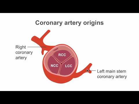
This detailed video from our Cardiac CT Essentials course will teach you how to locate the coronary artery origins with the help of cardiac CT. Dr Rajani will review the normal configuration of the right and left coronary arteries in relation to the aortic valve, explain the bifurcation and trifurcation anatomy of the left main stem, and offer an in-depth explanation of the differences between right and left coronary dominance.
Join our Cardiac CT Essentials course today!
Do you want to know how to perform and interpret your own cardiac CT scans? Our Cardiac CT Essentials course is designed and delivered by CT guru Dr Ronak Rajani. Consultant cardiologist, lecturer at King's College London, and Director of the London Advanced Cardiac CT Academy, Dr Rajani will be your comprehensive guide to the ins and outs of cardiac CT application in a clinical environment.

Become a great clinician with our video courses and workshops
Transcript
Coronary artery origins and left main stem anatomy
[0:00]
When we consider the origins of the coronary arteries, the normal configuration is that the right coronary artery arises from the right coronary cusp, while the left main stem arises from the left coronary cusp and then bifurcates into two separate arteries: the left anterior descending artery and also the left circumflex artery.
Left main coronary artery: bifurcation vs. trifurcation anatomy
[0:20]
The left main stem itself may adopt two main configurations. In left main stem bifurcation anatomy, the left main stem separates into the left anterior descending artery and also the left circumflex artery. In left main stem trifurcation anatomy, the left main stem separates into the left anterior descending artery and the left circumflex artery, but in addition there is a further branch, called either the ramus branch or the intermediate branch. This third branch normally follows a path between the left anterior descending artery and also the left circumflex artery. In this clip [lesson video], are you able to spot the trifurcation anatomy of the left main stem?
What coronary dominance means
[1:30]
When we describe the coronary arteries, the term that is commonly used is one called coronary dominance, and this can be an unnecessary source of confusion. Dominance of the right or left circumflex artery refers to the artery which gives off the posterior descending artery that supplies the inferior part of the heart. A patient is said to have right coronary dominance if this arises from the right coronary artery, and left coronary dominance if this arises from the left circumflex artery. Let us look at this concept a little bit more.
Right coronary dominance on CT
[2:02]
Here, we can see that the patient has right coronary dominance. The PDA that supplies the inferior aspect of the heart is supplied by the right coronary artery. It is worthwhile noting that up to 85% of hearts are right-sided dominant, and in these cases, the left circumflex artery is usually smaller.
Left coronary dominance and reporting it
[2:21]
Here, we can see that the patient has left coronary dominance, where the PDA supplying the inferior part of the heart is supplied by the left circumflex artery. A smaller portion of patients have left-sided coronary dominance, and when present, the right coronary artery is usually a small vessel. In clinical practice, the presence of coronary dominance is something to include in your reports, but is not usually something that is clinically relevant.
Right vs. left dominance: a quick check
[2:46]
This is an example of right coronary dominance. Can you see that the PDA arises from the right coronary artery? This is an example of left coronary dominance. Can you see that the PDA arises from the left circumflex artery? When issuing your CT reports, it is good practice to mention whether your patient has a right- or left-sided coronary dominant circulation.